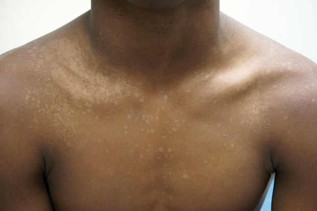
MKSAP Quiz: Recurrent skin eruption

A 25-year-old man is evaluated for recurrent skin eruption with oval lesions on the chest and upper back, which are occasionally itchy. The lesions began in late spring and worsened over the summer. Medical history is unremarkable, and he takes no medications.
On physical examination, vital signs are normal. Skin findings on the chest are shown.
Extremities and feet are unaffected.
Which of the following is the most likely diagnosis?
A. Candida albicans infection
B. Erythrasma
C. Pityriasis versicolor
D. Tinea corporis
Answer and critique
The correct answer is C. Pityriasis versicolor. This item is Question 2 in MKSAP 18's Dermatology section.
Pityriasis versicolor, often referred to as tinea versicolor, is one of the most common, chronic, superficial fungal infections. It is most common in warm, humid environments. It typically presents in young adults as asymptomatic, oval-to-round, minimally scaly, hyperpigmented or hypopigmented macules that can coalesce into patches on the trunk and upper extremities. Hypopigmentation is more common in darker-skinned persons. These areas often become more noticeable after exposure to the sun because the organism prevents the skin from tanning.
The diagnosis of pityriasis versicolor can often be made by clinical presentation but may be confirmed by visualization of short rod-shaped hyphae and round yeast (“spaghetti and meatballs”) on microscopic examination of skin scraping using a potassium hydroxide preparation. Treatment of pityriasis versicolor with topical antiseborrheic shampoos or lotions such as selenium sulfide or ketoconazole leads to resolution of erythema and scaling, but the pigmentation changes may persist for longer periods of time.
Candida albicans infections occur in hot, moist occluded areas, such as the armpits, groin, and beneath the breasts. Candida appears as erythematous patches with satellite pustules. Diagnosis is frequently made on clinical grounds alone, but a potassium hydroxide preparation can show spores and pseudohyphae.
Erythrasma is a superficial bacterial infection caused by Corynebacterium minutissimum that presents as mildly pruritic, thin erythematous-to-brown plaques with thin scale and an overlying wrinkled appearance and maceration in intertriginous areas such as the axillae, groin, and inframammary areas. The red-brown color and distribution in intertriginous areas distinguishes erythrasma from pityriasis versicolor. Diagnosis is typically made by clinical presentation but may be confirmed by coral-red fluorescence with a Wood lamp.
Tinea infections are superficial fungal infections caused by dermatophytes and classified by the site of infection. Tinea corporis appears in sites other than the feet, groin, face, or hand. It is characterized by annular patches with peripheral scale and central clearing that appear distinctly different from the hypo- or hyperpigmented patches of pityriasis versicolor. Diagnosis can often be made on clinical grounds, but a potassium hydroxide preparation of the scale shows fungal hyphae. A fungal culture can also be performed.
Key Point
- Pityriasis versicolor presents in young adults as asymptomatic, oval-to-round, minimally scaly, hyperpigmented or hypopigmented macules that can coalesce into patches on the trunk and upper extremities.


