MKSAP Quiz: 4-week history of joint pain, rash
A 33-year-old woman is evaluated for a 4-week history of recurrent worsening joint pain and a new rash on the chest and upper back. She reports sun exposure from spending time at the beach. She has no other symptoms. She has no other medical problems and takes no medications.

On physical examination, vital signs are normal. Diffuse tenderness of multiple small joints of the hands is noted. The remainder of the examination is normal.
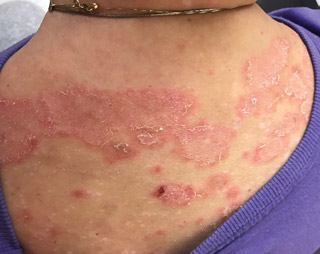
The appearance of the rash is shown.
Which of the following is the most likely diagnosis for the rash?
A. Acute cutaneous lupus erythematosus
B. Dermatomyositis
C. Discoid lupus erythematous
D. Subacute cutaneous lupus erythematous
Answer and critique
The correct answer is D. Subacute cutaneous lupus erythematous. This item is Question 93 in MKSAP 18's Rheumatology section.
This patient has subacute cutaneous lupus erythematosus (SCLE) and likely has systemic lupus erythematosus (SLE). She has a new rash on the torso with annular and patchy papular areas. This rash is most likely SCLE, a photosensitive rash occurring especially on the arms, neck, and upper trunk, usually sparing the central face. The rash can be annular with central clearing or papulosquamous with patchy erythematous plaques and papules. In some patients, the rash may have both of the characteristics, as seen in this patient. SCLE often has a fine scale that may leave postinflammatory hypo- or hyperpigmentation. SCLE is associated with anti-Ro/SSA antibodies, with a prevalence of 75%. About 50% of patients with SCLE also have SLE. These patients more typically have mild systemic symptoms, most commonly arthritis and myalgias; patients with lupus vasculitis, central nervous system lupus, and nephritis are found in less than 10% of patients with SCLE. Psoriasis can present with similar lesions but are usually photoresponsive (improve with sun exposure), unlike this patient.
Essentially 100% of patients with acute cutaneous lupus erythematosus (ACLE) have SLE. ACLE may present in multiple forms, with the most commonly recognized being a characteristic localized malar (butterfly) eruption. Less commonly it can appear as a generalized eruption, which typically appears as an erythematous maculopapular eruption of sun-exposed skin such as the extensor surfaces of the arms and hands.
Patients with dermatomyositis frequently have areas of poikiloderma with ill-defined patchy erythema and “salt-and-pepper” dyspigmentation accompanied by telangiectasias on the chest or upper back. There may be slightly violaceous erythema over the “V” of the neck, chest, and the upper back (the “Shawl sign”) or on the lateral hips (the “holster sign”).
Discoid lupus erythematosus (DLE) occurs in 20% of patients with SLE but more commonly occurs as an isolated, nonsystemic finding; patients with isolated DLE usually do not go on to develop SLE. DLE usually affects the scalp and face and presents as hypo- and/or hyperpigmented, possibly erythematous, patches or thin plaques that may be variably atrophic or hyperkeratotic.
Key Point
- Subacute cutaneous lupus erythematosus (SCLE) can present as annular with central clearing or papulosquamous with patchy erythematous plaques and papules, and both forms can be seen in the same patient; about 50% of patients with SCLE also have systemic lupus erythematosus.


