MKSAP Quiz: Gradually worsening dyspnea on exertion
A 32-year-old woman is evaluated for a 2-month history of gradually worsening dyspnea on exertion. She has difficulty climbing one flight of stairs, but has no wheezing or cough. She was hospitalized 6 months ago for a cholecystectomy complicated by sepsis, with a 2-week ICU stay requiring mechanical ventilation, but she recovered without difficulty. She uses an albuterol inhaler as needed, but this does not provide significant relief.

On physical examination, vital signs are normal. Oxygen saturation is 97% breathing ambient air. Cardiopulmonary examination is normal.
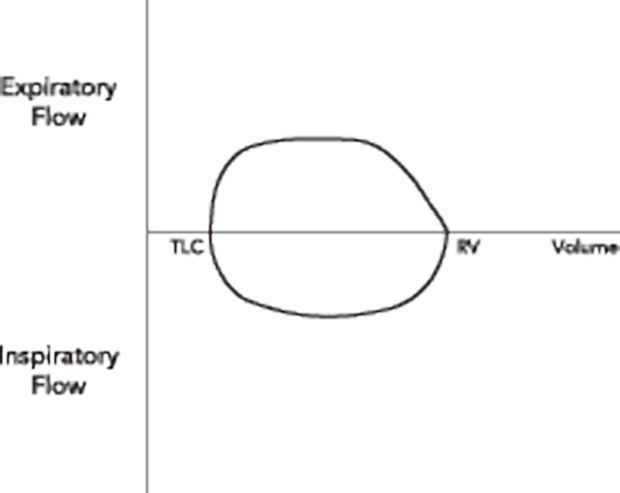
Chest radiograph is normal; spirometry is performed, and a flow-volume loop is shown.
Which of the following is the most likely diagnosis?
A. Asthma
B. COPD
C. Fixed upper airway obstruction
D. Normal lung function
Answer and critique
The correct answer is C. Fixed upper airway obstruction. This item is Question 89 in MKSAP 18's Pulmonology and Critical Care Medicine section.
The most likely diagnosis is fixed upper airway obstruction. Flow-volume loops graphically plot pulmonary airflow during exhalation and inspiration, with characteristic patterns associated with specific clinical conditions. The normal expiratory portion of the flow-volume loop (above the x-axis) is characterized by a rapid rise to the peak flow rate, followed by a nearly linear fall in flow as the patient exhales. The inspiratory curve (below the x-axis) appears as a semicircle. Flattening of both the inspiratory and expiratory curve of the flow-volume loop suggests this patient has a fixed intrathoracic lesion, such as tracheal stenosis, which can be seen postintubation. Spirometry is relatively insensitive to all but severe (greater than 71%) intrathoracic airway obstruction. Therefore, if intrathoracic airway obstruction is suspected, appearance of a normal flow-volume loop should not discourage further evaluation.
Patients with asthma would be expected to have this obstructive pattern, although patients without active symptoms can have a completely normal loop.
Patients with COPD demonstrate an obstructive pattern of the flow-volume loop. This is typically manifested by a fairly normal initial portion of the expiratory flow loop, with increased concavity of the terminal portion, indicating airway narrowing during exhalation.
The flow-volume loop demonstrates reduced inspiratory and expiratory volumes, and not the normal pattern described above.
Key Point
- Flattening of both the inspiratory and expiratory curve of the flow-volume loop suggests a fixed intrathoracic lesion; direct examination of the airways is indicated to confirm the finding and identify the cause.


