Treating skin ulcers in the office
In addition to five principles of wound care, an expert addresses pearls that can aid with all types of skin healing and antimicrobial efforts.
Internists should remember five main principles when it comes to treating wounds, said Theodore G. MacKinney, MD, MPH, FACP, a certified wound specialist physician: determining the need for debridement, assessing the need for antimicrobials, maintaining moisture, inspecting the edges, and detecting potential healing inhibitors.
“The key principle for wound care is to get to good wound-bed preparation. This is a technical term referring to a wound which has been optimized for healing,” he said. “It helps to have a systematic approach when looking at any wound, so looking at these five aspects is very helpful.”

First, inspect the wound for necrotic tissue and assess the need for debridement, said Dr. MacKinney. “Necrotic tissue promotes inflammation, which slows healing of chronic wounds, and it breeds infection,” he said.
That leads into the next principle: assessing the bacterial load and the need for antimicrobial therapy. “If there's an excess bacterial load, it also slows healing. This often presents with either increased pain or decreased healing of the wound,” said Dr. MacKinney, professor of medicine at Medical College of Wisconsin in Milwaukee.
Third, be sure to keep the wound moist, but not too wet or macerated. “We keep the wound from scabbing over because it heals faster and there will be less scarring that way,” he said.
Fourth, inspect the wound edges to see if they are heaped up or undermined. “This is often a problem with diabetic pressure sores and foot ulcers, and they need to be addressed surgically,” Dr. MacKinney said.
Finally, look outside the wound itself for potential inhibitors to healing. “Edema in the wound slows healing and is a large issue, especially with venous ulcers,” he said. “Venous and arterial circulation need to be addressed, as well as the control of diabetes.”
During his ACP CME 30 talk, Dr. MacKinney reviewed basic care for several types of wounds commonly seen in the outpatient setting, including diabetic foot ulcers and venous ulcers.
Diabetic foot ulcers
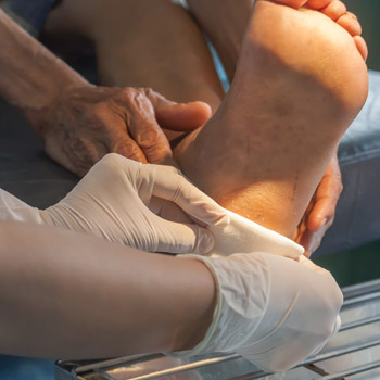
While it's already routine for internists to check the feet of patients with diabetes, Dr. MacKinney offered a deeper look at treatment and management of the wounded diabetic foot.
While the pooled prevalence of diabetic foot ulceration is about 6.3% worldwide, prevalence estimates in the U.S. and Canada are 13.0% and 14.8%, respectively, according to a systematic review and meta-analysis published in 2017 by Annals of Medicine.
“This issue is very important because two-thirds of all nontraumatic lower-extremity amputations are due to diabetic pressure ulcers,” said Dr. MacKinney. Other risk factors include soft-tissue infection and osteomyelitis, he noted. “Unfortunately, we're not making very good progress on this.”
He pointed to a study, published in 2019 by CMAJ, finding that lower-extremity amputations related to diabetes, peripheral artery disease (PAD), or both have been stable for 10 years in Canada. “There has not been a commensurate drop as there has been with cardiovascular disease and other complications of diabetes,” said Dr. MacKinney.
People with diabetes and lower health literacy may be especially at risk for foot amputation, he added. Compared to a general orthopedic patient population, those who had a diabetic foot amputation or reamputation in the last two years were eight times more likely to have inadequate health literacy, according to a study published in 2019 by the Journal of Foot & Ankle Surgery. “It reminds me that we need to give special attention to those who are more vulnerable,” said Dr. MacKinney.
He reviewed the care, dressings, and tests that are most useful for warding off infection and assessing those most at risk. When treating diabetic foot ulcers, make sure to check the patient's ankle brachial index (ABI) to assess for PAD, and glucose control should also be optimized, although “There is very little data that proves that this makes a difference,” he said.
These wounds often require sharp debriding, as well as pressure offloading, “which would include telling the patient not to walk with bare feet at all, even in their house,” Dr. MacKinney said, adding that patients typically require weekly visits by a nurse, podiatrist, or wound clinic staff during what can be a prolonged healing process.
For optimal healing, he recommended using topical antimicrobial dressings for all diabetic foot ulcers until the wound has been closed. Compared with nonantimicrobial dressings, antimicrobial dressings improve the rate of healing by about 30%, with 119 additional healing events per 1,000 participants, he said, citing a 2017 Cochrane review.
One antimicrobial dressing option is cadexomer iodine, which has been well tested in randomized controlled trials, Dr. MacKinney said. “This is actually a slow-release iodine that is better for the wound than using direct or diluted betadine in the wound,” he noted, adding that other acceptable choices include inexpensive, “old-school” silver sulfadiazine and polyhexamethylene biguanide, a nonspecific antimicrobial agent.
If soft-tissue infection spreads into the bone, patients can develop diabetic foot osteomyelitis. When assessing for osteomyelitis in the presence of a diabetic foot ulcer, the probe-to-bone test is a simple and effective tool, Dr. MacKinney noted. In a systematic review and meta-analysis published in 2016 by Clinical Infectious Diseases, the test had a sensitivity of 0.87 and a specificity of 0.83.
To help risk stratify patients with diabetic foot infection, new data point to the value of testing for procalcitonin, he noted. For example, a study of 86 inpatients, published in 2019 by the Journal of Diabetes Research, found that those with positive procalcitonin values (>0.5 ng/mL) had a 30.4% rate of limb salvage, compared to 93.6% among those without elevated values.
“A take-home message from this would be that if a patient has an elevated procalcitonin level, they should be treated more aggressively, earlier, and with more careful monitoring in hopes of salvaging the limb,” said Dr. MacKinney.
Finally, for patients with PAD and nonhealing diabetic foot ulcers, he recommended considering hyperbaric oxygen therapy. A systematic review and meta-analysis, published in February by the Journal of Vascular Surgery, found that the rate of major amputation in patients with diabetic foot ulcers and PAD was 25% among those who received standard treatment versus 10.7% among those who received adjuvant hyperbaric oxygen treatment, although there were no differences in wound healing, minor amputations, or mortality.
“I would say the data is still inconclusive, but I would refer patients who had nonhealing diabetic pressure ulcers and peripheral artery disease for hyperbaric oxygen therapy,” said Dr. MacKinney.
Venous ulcers
Venous ulcers are another type of wound you may see in the office. About 10% to 35% of U.S. adults will have venous insufficiency as they get older, and 4% of those over age 65 years are going to develop venous ulcers, Dr. MacKinney reported.
Risk factors for venous ulcers include older age, family history of ulcers and venous insufficiency, increased body mass, smoking, sedentary lifestyle, and ligament laxity, “where, if the patient has hernias and flat feet, they're more likely to have venous insufficiency and venous ulcers,” he said. Patients with venous disease often have a history of deep venous thrombosis (DVT) and, even if they don't, are three times more likely to have factor V Leiden mutation, Dr. MacKinney added.
The gold standard for diagnosing venous ulcers is a duplex scan, he said. “Every patient who has had a venous ulcer that has been at all difficult to treat should have a duplex scan to confirm the diagnosis,” said Dr. MacKinney, adding that 10% of leg ulcers occur from uncommon causes, including arterial ulcers and vasculitis, so a differential diagnosis should be considered. In addition, 2014 guidelines from the Society for Vascular Surgery and the American Venous Forum suggested evaluating for thrombophilia if there are recurrent ulcers.
While some primary care physicians may prescribe diuretics or oral antibiotics to treat venous ulcers, “Both of these are considered ineffective treatments,” said Dr. MacKinney. Instead, he said the main principles for treatment include local wound care and dressings, management of edema without using furosemide, and potentially surgery for recurrent or recalcitrant ulcers.
When assessing the wound, it's important to remove any offending substances, Dr. MacKinney said. “We need to remember that patients have likely put on something before they come and see us. Up to half of patients have contact dermatitis to topical treatment agents,” he said, adding that contact sensitivity is particularly common in patients with stasis dermatitis. Of these, “Up to 60% [have] dermatitis to common allergens, including lanolin, neomycin, parabens, and fragrances. We have even seen patients that are allergic to common cotton gauze,” Dr. MacKinney said.
For venous ulcers, no dressing has been proven better than another, he said, although “Absorbent dressings are often needed because the edema in the leg finds its way out of the wound, and these typically end up being very exudative wounds.” Therefore, using an alginate, foam, an ABD pad, or some other absorbent pad may be a necessary part of treatment. A 2012 Cochrane review found that pentoxifylline may help improve healing in some cases, Dr. MacKinney noted.
To reduce extremity edema, compression is the cornerstone of treatment. Multilayer compression dressings are the standard, he said, although Unna boots, support hose, and intermittent pneumatic compression devices may also be used. Other effective measures include elevation of the affected leg as much as is feasible for the patient, as well as exercise to help pump fluid and edema out of the lower leg, Dr. MacKinney said.
More pressure seems to be better. In one randomized study of 308 patients with recently healed venous ulcers, about 29% of those who received higher compression with class 3 compression stockings had ulcer recurrence after five years, compared to 60% of those who received class 2 compression, according to results published in 2018 by the Journal of Vascular Surgery–Venous and Lymphatic Disorders. “We would say more compression is better, but some compression is better than no compression,” he said.
Data comparing compression stockings and wraps are limited, but there is a consensus that multilayer wraps are more useful for initial edema reduction and wound management, Dr. MacKinney said. That's because a Jobst stocking or other hosiery will be measured according to the size of the edematous leg, and when the swelling goes down, it won't compress the leg as well anymore, he noted. “It is better to get all of the edema out of the leg with compression dressings, and then, when the wound is healed, to use stockings for maintenance,” said Dr. MacKinney.
There are many types of wraps that can be used, including short-stretch bandage-based systems, no-stretch options (e.g., Unna boots), and long-stretch bandages like Ace bandages. “The preferred modality for wound clinics is to use short-stretch bandage systems,” which provide lower pressure at rest but higher working pressure during exercise, rather than the high but fixed pressure of long-stretch bandages, he noted. “It's these large increases in pressure which pump fluid out of the leg.”
Compression dressings should not be used if there is an acute infection or cellulitis in the leg, and there's also a relative contraindication if the ABI is less than 0.8, Dr. MacKinney said. They should also be avoided if there is uncompensated congestive heart failure, he said, “because you dump a large amount of proteinaceous fluid into the vascular system, which may precipitate pulmonary edema.”
In addition, do not use compression in the setting of acute thrombophlebitis; wait about two weeks to start compressing the leg after a DVT, said Dr. MacKinney. Finally, he recommended using caution if there's neuropathy in the leg because there may be a compression injury that the patient doesn't know about.
Venous ulcers can take a long time to heal and may also recur. They are slower to heal if the patient is older, if the wound has been there a long time, or if the wound is large, Dr. MacKinney noted. An ulcer is more likely to recur if the original healing was slow or if the superficial reflux system of the veins was not treated with surgery, he said.
That's why clinicians should consider referring patients with recurrent venous ulcers for early surgery, Dr. MacKinney said. In the EVRA (Early Venous Reflux Ablation) trial, participants randomized to early endovenous ablation of superficial venous reflux within two weeks had a median time to ulcer healing of 56 days, compared to 82 days for those who waited up to six months to have the surgery, according to results published in 2018 by the New England Journal of Medicine.
“A later assessment of the data also showed that there is improved quality of life at one year for those that have been operated on,” said Dr. MacKinney. “It's my impression that even in our wound clinics, venous ulcers should be seen sooner by surgeons than they are right now.”


