MKSAP Quiz: 8-year history of joint pain
A 50-year-old man is evaluated for an 8-year history of joint pain, particularly in the hands, and progressively worsening fatigue. Medical history is otherwise unremarkable. He takes ibuprofen as needed.

On physical examination, vital signs are normal. BMI is 32. There are swelling and tenderness of the second and third metacarpophalangeal (MCP) joints bilaterally, and tenderness but no swelling of the fourth and fifth MCP joints bilaterally. There is bony hypertrophy of the first MCP joints and knees bilaterally. The proximal interphalangeal joints and wrists are normal.
Laboratory studies are notable for an alanine aminotransferase level of 53 U/L and an aspartate aminotransferase level of 55 U/L; rheumatoid factor is negative.
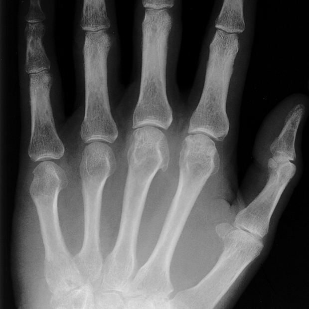
A radiograph of the hand is shown.
Which of the following is the most appropriate diagnostic test to perform next?
A. Anti–cyclic citrullinated peptide antibody assay
B. Hepatitis C antibody assay
C. Serum α-fetoprotein measurement
D. Transferrin saturation measurement
Answer and critique
The correct answer is D: Transferrin saturation measurement. This question can be found in MKSAP 17 in the Rheumatology section, item 11.
Measurement of transferrin saturation is the most appropriate diagnostic test to perform next in this patient. He has signs and symptoms suggestive of hemochromatosis, an autosomal recessive disorder characterized by increased absorption of iron from the gut. Approximately 40% to 60% of patients with hemochromatosis develop arthropathy that is osteoarthritis-like, but characteristically involves the second and third metacarpophalangeal (MCP) or wrist joints. Transferrin saturation and serum ferritin levels are usually elevated in patients with arthropathy due to hemochromatosis. Although a variety of more specific diagnostic maneuvers may be undertaken, including liver biopsy and genetic testing for homozygosity for the C282Y mutation of the HFE gene, the most appropriate and cost-effective next step is measurement of transferrin saturation. A consensus does not exist for transferrin saturation cut-off levels for diagnosis of hemochromatosis, with some guidelines recommending a value of greater than 60% in men or greater than 50% in women, and others suggesting a level of greater than 55% for all patients. Measurement of ferritin levels is indicated in patients with an elevated transferrin saturation; a markedly elevated level further supports the diagnosis and predicts the development of symptoms. The presence of clinical MCP involvement with radiographic evidence of hook-shaped osteophytes is most characteristic of hemochromatosis.
Anti–cyclic citrullinated peptide antibodies are never associated with hemochromatosis, and rheumatoid factor is generally negative in patients with hemochromatosis as seen in this patient. These autoantibodies have specificity for rheumatoid arthritis.
Arthritis may occur in up to 20% of patients with hepatitis C virus infection and may mimic rheumatoid arthritis clinically and radiographically. However, the characteristic findings of hemochromatosis on this patient's radiographs and the lack of more typical findings of erosions or bony decalcification adjacent to the involved joints make a diagnosis of hepatitis C–associated arthritis less likely.
Serum α-fetoprotein is elevated in liver disease such as acute or chronic viral hepatitis infection as well as in hepatocellular and numerous other cancers. It would have little diagnostic specificity in this clinical setting.
Key Point
- Secondary osteoarthritis may occur in the setting of hemochromatosis, which is associated with an arthropathy that is osteoarthritis-like, but characteristically involves the metacarpophalangeal and wrist joints.


