MKSAP Quiz: Evaluated in the ED after a fall
A 66-year-old man is evaluated in the emergency department following a fall 3 days ago, when he struck the right side of his chest against a table. Since that time he has experienced right-sided pleuritic chest pain and difficulty taking a deep breath. His only other medical problem is chronic atrial fibrillation, for which he takes warfarin.
On physical examination, temperature is 37.3°C (99.1°F), blood pressure is 132/80 mm Hg, pulse rate is 75/min and irregular, and respiration rate is 15/min with significant left-side splinting; BMI is 26. Oxygen saturation is 85% breathing ambient air. The right side of his chest shows extensive bruising. There is no jugular venous distention and, other than an irregularly irregular rhythm, the cardiac examination is unremarkable. There are diminished breath sounds over the right lower chest with occasional crackles.
Oxygen saturation does not improve with increasing flow rates of supplemental oxygen delivered by nasal cannula. Oxygen saturation of 90% is achieved with 80% supplemental oxygen delivered by mask.
Laboratory studies reveal a hemoglobin level of 11.8 g/dL (118 g/L) and an INR of 2.5.
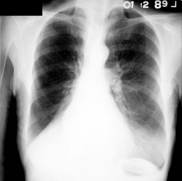
Chest radiograph is shown.

Which of the following is the most likely diagnosis?
A. Hepatopulmonary syndrome
B. Intracardiac shunt
C. Intrapulmonary shunt
D. Pulmonary arteriovenous malformation
Answer and critique
The correct answer is C: Intrapulmonary shunt. This question can be found in MKSAP 17 in the Pulmonary & Critical Care Medicine section, item 53.
The most likely diagnosis is intrapulmonary shunt. This patient's profound hypoxia is most likely due to intrapulmonary shunting of blood through his collapsed right lower lobe. Evidence of a collapsed right lower lobe is indicated by the triangular opacity above the diaphragm on the right side on his chest radiograph. Right-to-left intrapulmonary shunts may be anatomical or physiological, but all result in hypoxemia that does not readily correct with supplemental oxygen, as seen in this patient. This patient's physiological shunt results in perfusion of nonventilated alveoli of the collapsed right lower lobe. In most situations, a lobar collapse will not cause such severe shunting because of hypoxic vasoconstriction of arterioles in parts of the lung that are not ventilated; however, in some cases, such as this patient, this compensatory mechanism directing blood flow to other aerated areas of the lung is ineffective.
Hepatopulmonary syndrome, intracardiac shunt, and pulmonary arteriovenous malformation are all examples of anatomic shunts. Although these conditions can be responsible for hypoxemia that is difficult to correct with supplemental oxygen, this patient's trauma, splinting, and abnormal radiographic findings suggest a lower lobe collapse as the most likely diagnosis. In conditions of diagnostic uncertainty, a bubble contrast echocardiogram could be performed. These anatomic shunts are associated with the appearance of left atrial or ventricular bubbles after several cardiac cycles.
Hepatopulmonary syndrome should be suspected in patients with cirrhosis who develop hypoxemia in the absence of other causes. Their shunt worsens with upright posture (orthodeoxia). However, this is not a likely diagnosis in a patient without evidence of cirrhosis.
Key Point
- Right-to-left intrapulmonary shunts may be anatomic or physiologic, but all result in hypoxemia that is difficult to correct with supplemental oxygen.


