Know your kidney stones
Knowing what makes up a patient's kidney stones determines how an internist might treat the problem. But, one expert cautions, while 1 type of kidney stone is far more prevalent, there are 4 other kinds, and they may require subspecialty treatment.
Kidney stone treatment and prevention depend on the type of stone you're dealing with, according to Gary C. Curhan, MD, ScD.
“When I see a patient I always ask them what type of stone they had, and usually they raise their voice a little bit and say, ‘A kidney stone,’ as if I didn't hear them the first time,” he said. “But what you and I really want to know is what the stone's made out of.”

Calcium oxalate is the most common type of stone, seen in 74% of first-time stone formers and 66% of recurrent stone formers. “Overall, if you are not sure of the stone composition, you should always guess calcium oxalate, far and away,” said Dr. Curhan, who is a professor of medicine at Harvard Medical School and a member of the Renal Division at the Brigham and Women's Hospital in Boston.
Most of Dr. Curhan's talk pertained to calcium oxalate stones, but he also mentioned 4 other types, 2 of which, cystine and struvite, should always indicate referral to a subspecialist. Cystine stones are caused by an autosomal recessive disorder and are unrelated to diet. They are evaluated by measuring 24-hour cystine excretion and prevented by using tiopronin or penicillamine and raising urine pH.
“There's nothing that we can do to change the amount of cystine that's coming out. We don't put people on low-cystine diets because that just won't work. So we do things to try to change the solubility, and it can be quite effective,” said Dr. Curhan.
Struvite stones, also called “infection stones” or Mg-NH4+ carbonate-apatite stones, only form when urease-producing bacteria are present in the upper urinary tract, Dr. Curhan noted. Complete stone removal is required, and further stones can be prevented by preventing urinary tract infections (UTIs), he said.
Dr. Curhan said that calcium phosphate stones are his least favorite type because they are difficult to prevent. They are caused by too much calcium in the urine, too much phosphate in the urine, or too little citrate in the urine. These stones only form when the urine is more alkaline, which can occur with certain bacteria or renal tubular acidosis. They can be prevented by thiazide and citrate, although the latter may raise pH and increase calcium phosphate stone formation, Dr. Curhan said.
In contrast, uric acid stones are very easy to prevent, Dr. Curhan said. They are usually caused by low urine pH and sometimes by elevated urine uric acid. Prevention involves decreased intake of animal protein, which reduces purine consumption and acid generation. Alkalinization of urine to a pH of 6.5 to 7.0 and xanthine oxidase inhibitors can also help, Dr. Curhan noted.
Causes and risk factors
Nephrolithiasis has some systemic contributors, such as primary hyperparathyroidism. “We always look for this because it's one of the few curable causes of stone formation,” Dr. Curhan said. “These people have high serum calcium and at least a high urine calcium.”
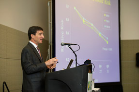
Increasing weight also probably plays a role in the increase in stone disease that's been seen in the U.S., Dr. Curhan said. He pointed to data from the Health Professionals Follow-up Study, the Nurses' Health Study I, and the Nurses' Health Study II showing that women who weighed 220 pounds or more had an 80% higher risk of stone formation than those who weighed less than 150 pounds; in men, there was a 40% higher risk.
“Unfortunately we don't have enough people who lost weight that I can tell you that losing weight would be protective, but I can at least say that weight maintenance is essential,” he said.
Urinary risk factors for kidney stones include high concentrations of calcium and citrate, while higher citrate and higher urine volume confer lower risk, Dr. Curhan said. Urine uric acid was previously thought to be a risk factor for calcium oxalate stones, but that's not the case, he noted.
Evaluation
Dr. Curhan doesn't use terms like hypercalciuria, hyperoxaluria, or hypocitraturia in evaluating patients. “Those are dichotomous terms, and I think they've really held the field back because people say ‘Oh, it's abnormal’ or ‘It's normal,’ and these are really continuous variables,” he said. “The analogy I use all the time is that this is like blood pressure. We may say that someone has hypertension if their blood pressure is 141 over 91 [mm Hg], but if they're 139 over 89, everything isn't fine.”
Also, he said, the more extreme the numbers, the higher the risk. “So to say, ‘They have hypercalciuria,’ I want to know what the number is. Is it 200, or is it 600? Because those things are really going to make a big difference in the likelihood of crystal formation.”
Traditional reference ranges for hypercalciuria, hyperoxaluria, and hyperuricosuria are also not helpful, Dr. Curhan said. “These are not based on normal distribution. It's just somebody at some point said ‘That's too high,’” he said. “It isn't like serum sodium or serum potassium. The definition for hyperoxaluria is arbitrary, and the definition for hyperuricosuria as well. So again, think about these more continuously.”
When examining a patient with a first-time kidney stone, clinicians should make sure to look for other residual stones on imaging studies that were done, Dr. Curhan said.
“If they have other stones in their kidney, to me they're a recurrent stone former. They just aren't a recurrent stone passer yet,” he said.
Evaluation is recommended not only for patients with residual stones but also those at high risk for recurrence, said Dr. Curhan. It's also recommended in kidney stone patients with osteopenia or osteoporosis. “That's another reason why we'd want to know how much urine calcium is coming out and whether we should do something about it,” he noted.
The initial step in the evaluation is the history, specifically focusing on the number of stones, the frequency, and the types of interventions taken in response. “These are all things [that] the higher they are, the more likely I am to be aggressive to reduce the values,” Dr. Curhan said.
Electrolytes, renal function, calcium, and phosphorus will usually have been checked before you see the patient, Dr. Curhan said. “I don't check PTH, or parathyroid hormone, when I first see somebody, unless there's evidence of either their urine calcium being high—but usually they haven't had it checked before they've come to see me—or if their serum calcium is high,” he said.
In the acute setting, a spot urinalysis should be done to be sure the urine isn't infected, Dr. Curhan stressed, since this would indicate a medical emergency in the presence of an obstructing stone. “People aren't going to die from the pain [of a kidney stone], but if there's an obstructing stone with an infection behind it, that is a situation where you need emergent decompression. And people do die from this,” he said.
Also, he noted, crystals in the urine of a patient who hasn't passed a stone yet or didn't retrieve the stone when it passed can help the clinician determine the stone type.
The 24-hour urine test is the cornerstone of recommendations, Dr. Curhan said, but he noted that it should not be performed until at least 6 weeks after the initial presentation.
“After someone's had any medical event, but particularly a kidney stone, they start changing everything that they do. But I don't want them to change their habits yet because I don't know what they need to change or not, because I don't know when that stone formed,” he said. He advises people to go back on their usual diet, wait at least 6 weeks, and then do two 24-hour urine collections for a baseline because excretion can vary, he said.
It's also very important to measure everything at once, he stressed. “Don't just measure the calcium and say ‘Oh, that's OK,’ and then have them do a 24-hour urine and measure the oxalate and that's OK. If you've never done a 24-hour urine and you're going to order it, I would have you try to do [what your patient will have to do]. Conceptually it's easy, but it can be challenging.”
For radiology, low-dose spiral CT is the standard of care, he said. KUB (kidneys, ureters, bladder) imaging uses much less radiation but can miss small stones. Ultrasound only visualizes the kidney and can't distinguish if there are 2 stones next to each other, but it has the benefit of not involving any radiation, he said.
Interventions
Dr. Curhan also summarized the pharmacologic interventions he uses for kidney stones. To try to reduce urine calcium, he would start with chlorthalidone, at least 25 mg/d, although he noted that some patients may eventually need a dose as high as 100 mg/d. “If you prefer hydrochlorothiazide, you may have to give it [with twice-daily] dosing just because the duration of action is shorter,” he said.
To try to reduce uric acid or decrease urine citrate, prescribe allopurinol or potassium citrate, respectively, he said. Any type of alkali will lead to increased citrate excretion, he noted, but he said he tries to avoid the sodium salts because higher sodium intake increases calcium excretion.
Patients taking thiazides must follow a low-sodium diet, he stressed, preferably less than 3 g/d. Actual intake should be quantified in 24-hour urine. “I can't tell you how many people say they do not eat any sodium, and you measure the 24-hour urine sodium and it's 4,000 mg/d,” he said. “The body does not make sodium. If they're in a steady state, that has to be coming from their foods.”
The challenge, he noted, is that prepared foods have unknown sodium content and labels can be tricky to read. “Just try to make patients aware of sodium intake,” he said.
Dr. Curhan also discussed dietary interventions to prevent stone formation. Dietary oxalate is probably not a huge contributor for most patients, but patients should try to avoid high-oxalate food such as spinach and potatoes, he said. Also, he noted that increasing dietary calcium has actually been shown to lower the risk of kidney stones. “There's no evidence at all to support severe calcium restriction in people that have calcium stones. I can't emphasize that enough,” he said.
Calcium supplements may be linked to kidney stone formation because of the timing of ingestion, he said. “Most people don't take their calcium supplements with meals; they usually take them in the morning or late at night,” he said.
Fluid intake is essential to prevent stone recurrence, Dr. Curhan stressed. “This is a disease of concentration,” he said. “Many times I get patients sent to me and the [referring clinician will] say ‘All their values in the urine are normal. Why are they forming stones?’ But then you collect a 24-hour urine and in fact their urine volume is so low that the concentration of those constituents [is] just too high.”
When patients are encouraged to increase fluid intake, they may ask what they can drink, Dr. Curhan said. Data show that higher intake of coffee, tea, beer, wine, and orange juice is associated with lower risk of stone formation, Dr. Curhan said. Tea does have oxalate in it but it's not a high amount, he said, and observational data show that there's no reason to avoid it.
“I do want to point out that I don't prescribe beer and wine to prevent kidney stones,” he joked, but he noted that it can help patients to know that drinking these beverages doesn't increase their risk. “I do advise them to avoid sugared sweetened beverages because those have been associated with a high risk of stone formation in addition to all their other adverse effects.”
Dr. Curhan acknowledged that it can be hard for patients, and even clinicians, to make sense of specific nutrient advice and that a general dietary approach can be easier. He pointed out that 3 observational cohorts have found that the DASH (Dietary Approaches to Stop Hypertension) diet, which is rich in fruits and vegetables, includes low-fat dairy, and has low amounts of saturated and total fat, is associated with lower risk. “The more DASH-like your diet, the lower risk of forming a stone,” he said. He also said that dietary recommendations should be individualized based on each patient's urine chemistries.
Several factors should be considered when choosing between dietary or pharmacologic interventions, Dr. Curhan said, including stone composition, stone-forming activity, previous procedures, urinary values, and bone density. For a woman with more extreme urinary values and low bone density, for example, he is more likely to prescribe medication. Patient preferences also play an important role.
“[A patient may say] ‘How long do I have to take this for?’ The real answer is ‘Forever,’ but what I usually say is ‘Well, we're not curing anything. It's not like a week of antibiotics. Let's try it for a year and see how it goes,’” Dr. Curhan said. “And usually, if you can prevent new stones, they're so happy that they're willing to continue it. But if they say they won't do it for a long term, it isn't worth starting them on medication.”
Performing the initial 24-hour urine measurement and advising the patient on next steps are only the beginning where kidney stones are concerned, Dr. Curhan said. To prescribe a thiazide and tell the patient to come back if he has another stone is “like treating someone's blood pressure and saying ‘Come back if you have a stroke,’” he said. “We would never do that. We measure people's blood pressure because we want to see what's happening.”
The 24-hour urine measurements should be repeated to see the effect of the medications prescribed, Dr. Curhan stressed. For example, you can see whether urine calcium decreased after a thiazide was started or whether urine sodium decreased after you recommended a low-salt diet.
“You may have to go through this loop just a couple of times,” he said, “but eventually you can dramatically change the composition of the urine so that stone recurrence will stop.”


