MKSAP Quiz: 4-day history of fever, malaise
A 24-year-old man is admitted to the hospital with a 4-day history of fever, malaise, and arthralgia of the elbows, wrists, and knees. Two days ago, he developed progressive pain and swelling of the right knee. He also has a rash on his right arm.
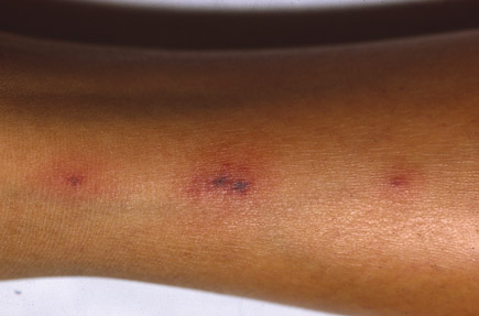
On physical examination, temperature is 38.2°C (100.8°F), blood pressure is 110/60 mm Hg, pulse rate is 95/min, and respiration rate is 12/min. There is evidence of tenosynovitis of the left wrist. The right knee is swollen and warm, with significant effusion. The rash on his arm is shown.

Blood cultures are obtained, and an arthrocentesis of the right knee is performed. The synovial fluid leukocyte count is 60,000/µL (60 × 109/L) with 90% polymorphonuclear neutrophils. The Gram stain is negative.
Which of the following is the most appropriate diagnostic test to perform next?
A. Antinuclear antibody and rheumatoid factor assays
B. Biopsy and culture of a skin lesion
C. HLA B27 testing
D. Nucleic acid amplification urine test for Neisseria gonorrhoeae
Answer and critique
The correct answer is D: Nucleic acid amplification urine test for Neisseria gonorrhoeae. This question can be found in MKSAP 16 in the Infectious Disease section, item 29.
The most appropriate next step in diagnosis is a nucleic acid amplification urine test for Neisseria gonorrhoeae. This is a noninvasive, sensitive test for diagnosing gonorrhea in men that provides rapid results (within hours) and can help guide therapy pending return of blood and synovial fluid culture results. Mucosal cultures, including of the throat, anus, urethra, or cervix, may also be helpful in establishing the diagnosis because they tend to have a higher diagnostic yield than blood and synovial fluid cultures in patients with disseminated gonococcal infection (DGI).
Although this young patient may have an autoimmune inflammatory arthritis such as systemic lupus erythematosus or rheumatoid arthritis, he has evidence of an arthritis-dermatitis syndrome and should be evaluated for DGI. In contrast to nongonococcal septic arthritis, patients with DGI present with migratory joint symptoms and often have involvement of several joints with tenosynovitis rather than involvement of just a single joint. Asymmetric joint involvement helps distinguish DGI from autoimmune disease associated polyarthritis, which is typically symmetric. Skin lesions are found in more than 75% of patients with DGI but may be few in number; consequently, a careful examination of the skin must be performed. Lesions are most likely to be found on the extremities. The classic lesion is characterized by a small number of necrotic vesicopustules on an erythematous base. Organisms are rarely cultured from the skin lesions of DGI, although they may be demonstrated through nucleic acid amplification techniques.
The prevalence of HLA B27 in patients with reactive arthritis is only 50%; consequently, HLA B27 testing is not very useful in establishing a diagnosis. In addition, reactive arthritis tends to present as an asymmetric oligoarthritis, and this patient's arthritis is monoarticular. The associated rash, keratoderma blennorrhagica, consists of hyperkeratotic lesions on the palms and soles, which are not present in this patient. Patients with reactive arthritis may also have conjunctivitis, urethritis, oral ulcers, and circinate balanitis.
Key Point
- To confirm a presumptive diagnosis of disseminated gonococcal infection, in addition to blood and synovial fluid cultures, specimens should be obtained from mucosal surfaces, including the throat, anus, and urethra, or cervix, which can be tested via nucleic acid amplification tests or culture.


