MKSAP Quiz: 5-day history of asymptomatic rash
A 68-year-old man is evaluated in the emergency department for a 5-day history of an asymptomatic rash on the upper back and upper arms. The lesions grew suddenly and are neither pruritic nor painful. The patient has had associated fevers. He has a myelodysplastic syndrome and has received intermittent erythrocyte transfusions, and he takes azacitidine.
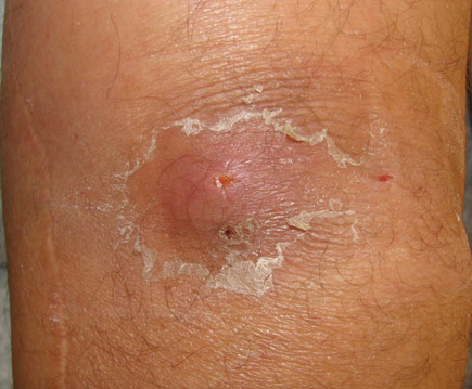
On physical examination, temperature is 39.0°C (102.0°F), blood pressure is 140/76 mm Hg, pulse rate is 109/min, and respiration rate is 20/min. The patient is pale and slightly ill appearing. There is conjunctival rim pallor and scattered petechiae with ecchymoses of various stages. There are nodular and plaque lesions on the back and upper arms. An illustrative example is shown.

Skin biopsy reveals a dense neutrophilic infiltrate throughout the dermis, with prominent papillary dermal edema.
Laboratory studies are as follows:
| Hemoglobin: | 7.2 g/dL (72 g/L) |
| Leukocyte count: | 35,000/µL (35 × 109/L) neutrophil predominance, circulating blasts |
| Platelet count: | 32,000/µL (32 × 109/L) |
Which of the following is the most likely diagnosis?
A. Herpes simplex virus infection
B. Leukemia cutis
C. Methicillin-resistant Staphylococcus aureus abscess
D. Pyogenic granuloma
E. Sweet syndrome
Answer and critique
The correct answer is E: Sweet syndrome. This question can be found in MKSAP 16 in the Dermatology section, item 34.
Sweet syndrome is a neutrophilic dermatosis that can be seen as a reaction pattern associated with underlying malignancy, particularly acute myeloid leukemia. In patients with myelodysplastic syndrome, development of Sweet syndrome may coincide with transformation to acute myeloid leukemia. This patient has classic clinical morphology, with bright red plaques, well demarcated with a sharp cut-off separating normal and inflamed skin. Because of the intense neutrophilic inflammatory infiltrate and accompanying papillary dermal edema, the lesions often look “juicy” and in some patients may mimic incipient bullae. Patients with Sweet syndrome are frequently neutrophilic and almost always have fevers.
Herpes simplex virus infections can present in numerous ways in immunocompromised hosts; however, there are nearly always signs of cutaneous vesicles that may rupture and cause erosions. Bilateral nodular lesions would be extremely atypical.
Leukemia cutis may present in multiple ways, but traditionally lesions are red-to-violaceous dermal papules and nodules. A biopsy of leukemia cutis would reveal atypical, leukemic cells infiltrating the dermis and dissecting through the collagen bundles.
Methicillin-resistant Staphylococcus aureus abscesses often present with large, indurated, red, tender, warm plaques, but there is almost always a crusted papule, pustule, or “head” at the center of the furuncle, which is lacking in this patient.
Pyogenic granulomas are small benign vascular papules with a collarette of scale, which can occur almost anywhere but tend to be seen on extremities around the nails and on the face. Patients on highly active antiretroviral treatment or certain acne medications or pregnant women may be more prone to develop these lesions.
Key Point
- Typical lesions of Sweet syndrome are “juicy,” bright red, well-demarcated plaques with a sharp cut-off separating normal and inflamed skin that appear on the neck, upper trunk, and extremities.


