Learning to learn: Ophthalmoscopic skills taught by simulation
Creating an ophthalmoscopic examination simulation model is easy, inexpensive and goes a long way toward improving this basic clinical skill set. Acquaint yourself with abnormalities seen in the office, as well as the motor skills involved with proper use of the instruments.
When learning or updating ophthalmoscopic skills, it can often be more effective to provide a simulation experience that more closely mimics a real-life examination. Not only can learners acquaint themselves with common ophthalmologic abnormalities that they will encounter in the office, but they can practice the motor skills involved with proper ophthalmoscope use.
To tackle both of these challenges, ACP uses an inexpensive ophthalmoscopic simulator for workshops held each year at the Internal Medicine meeting. The model was first developed by Edward A. Jaeger, MD, who is professor of ophthalmology and attending surgeon in the department of ophthalmology at Jefferson Medical College and Wills Eye Institute in Philadelphia. With the right teaching materials and a minimum of equipment, this skill can be easily taught in the institutional or office setting.
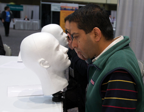
Creating an ophthalmoscopic examination simulation model is easy, relatively inexpensive and can provide learners the practice opportunity they need to improve this basic skill. The following is a step-by-step process for creating a homemade eye exam model, the equipment needed for the procedure, and a basic guide for putting together a workshop on the topic.
Creating the simulation model
Hardware needed:
- 1 Styrofoam mannequin head/wig stand. Styrofoam wig stands can be found online at numerous sites that sell professional beauty supplies and equipment for hair stylists. The heads usually range from $10-$25 per piece. Be sure to choose a head that is as anatomically correct as possible. Stylized wig stands that do not realistically represent the human head may not provide the proper structure to hold the portable slide viewer as needed.
- 1 marker or pen
- 1 power drill (with ¼- and ½-inch drill bits)
- 1 sharp utility blade or kitchen knife
- 1 sharp hobby knife (X-Acto or similar brand) or scalpel
- 1 mannequin head table clamp stand. The table clamp stands are also available from websites that specialize in professional beauty and hair styling supplies. The stands usually range from $6-$24 per piece.
- 1 backlit pocket slide viewer with batteries (Kalt, Omega, or Arista make models that are small, flat, and suitable for using in this simulation. They usually range from $4-$9 each.)
- 35-mm slides of normal and abnormal ophthalmologic findings
Instructions:
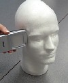
Step 1a. Turn the portable slide viewer on its side and hold up to one side of the mannequin head level with the eye. Make sure that the eye of the mannequin head is centered between the top and bottom edges of the slide viewer.
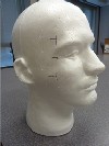
Step 1b. Use a marker or pen to mark the top and bottom edges of the slide viewer on the side of the head.
Step 2. Using the drill and 1/2-inch bit, drill about 2.5 to 3 inches straight into the side of the mannequin head below your top mark, and again above your bottom mark.
Be careful not to angle the drill bit, as you want to create a space behind the mannequin eye that is as exactly perpendicular to the side of the head as possible. You may also want to mark your drill bit at the 2.5- to 3-inch mark, so you know you are going in far enough and not going too far.
Step 3. Using the same method, drill out the rest of the slot in between the first two holes. Use the sharp utility blade or kitchen knife to cut away any rough parts inside the slot and generally smooth out the edges of the slot.

Step 4. Using the 1/4-inch drill bit, drill a hole through the center of the mannequin eye until you break through into the slot you've made.
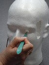
Step 5. Using the hobby knife or scalpel, carefully shave away the inside of the eyehole so that it opens up as you move into the slot for the slide viewer. From the entrance of the eye you should have a roughly cone-shaped opening. Shaping the eye opening like this is important as it will allow learners using an ophthalmoscope to look up and down, left and right at the slide image and mimic the motor skill of viewing the different quadrants of the eye chamber.
Step 6. Clamp the mannequin head stand to the edge of a table and mount the mannequin head on the adjustable pole.
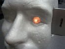
Step 7. Insert a 35-mm slide into the slot in the proper orientation, turn on the back light and slide the portable slide viewer into the newly cut slot. Make sure the slot is deep enough, at a straight angle, and that the image of the eye is centered in front of the eye opening.
About the slide images
You will need good-quality images of ophthalmological abnormalities in order to properly use this simulation model. Many institutions will have access to images via libraries or from teaching physicians. If you are recruiting an ophthalmologist to teach a workshop, you can ask the faculty to provide his or her own images.
Care should be taken to make sure the images are roughly the same size and not cropped too much by the edges of the 35-mm slide. Images should be of a high enough resolution that smaller images can be resized without getting pixilated.
It is best to get electronic files of the images, so that they can be resized, brightened, darkened, or otherwise manipulated before being made into 35-mm slides. If you do not have access to a graphic service in your institution or office setting, there are numerous websites that will make 35-mm slides of uploaded files. (Websites such as PhotoGraphic Specialties, Express Slides, or Slideplus all offer quick online solutions.)
Suggested teaching format
A typical ACP Clinical Skills workshop will consist of didactic background content, some faculty demonstration, and ample time for participants to practice the procedure or examination skill with feedback from the faculty.
A suggested format for a 90-minute to 2-hour workshop would be as follows:
- 30-40 minutes of didactic content (eye anatomy, review of direct ophthalmoscopy, common eye diseases)
- 20 minutes of practice with ophthalmoscopes (participants can practice on each other for this portion of the workshop)
- 30-60 minutes viewing slides of ophthalmologic abnormalities and discussion. It helps to have background diagnostic information of each condition or disease printed out and stationed next to the simulator. Participants can review this information as they examine the eye with the scope.
(Note: You will need access to ophthalmoscopes for this workshop, and should have one scope for every two participants.)
There will be two opportunities for hand-on ophthalmoscopic skills learning at the 2011 Internal Medicine Meeting in San Diego, on April 7-9. There will be a reserved workshop on Thursday, April 7 at 7 a.m. in room 15A. There will also be an open-access self-study area in the Herbert S. Waxman Clinical Skills Center, located in Hall A of the convention center. The self-study activity will run during all three days of the meeting and does not require a ticket. Participation is on a first-come, first-served basis. More information on is online.

