Spotting thyroid nodules entails history—of patient and art
An art masterpiece inspired goiters as a fashion trend in Baroque-era Europe, and in the modern age kicks off a discussion on diagnosing nodules that can be palpated in only 7% of women and 2% of men. An experts counsels how to detect them.
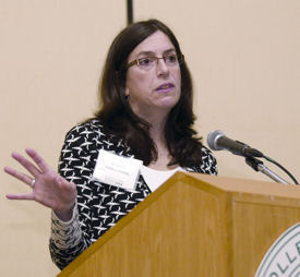
When Peter Paul Rubens painted Marie de Medici with a goiter in 1622, it set something of a trend. All the gentlewomen of the day insisted goiters be added to their own portraits as well—even if they didn't have them in real life, noted Susan Mandel, FACP, at the start of her talk on thyroid nodules at Internal Medicine 2009.
Goiters are still common—if not fashionable—in parts of the world where iodine deficiency is endemic, but they haven't been seen as much in the U.S. since the introduction of iodized salt in the 1920s. Still, thyroid nodules are fairly prevalent. The average American has a 50% chance of a thyroid nodule being detected on ultrasound by the time he or she reaches age 60, said Dr. Mandel, Professor of Medicine and Radiology at the University of Pennsylvania in Philadelphia.

“Interestingly, only about 7% of those nodules in women, and 2% in men, would be detected by palpation,” Dr. Mandel said.
The good news is that 90-95% of thyroid nodules are benign. But how to detect the other 5-10%?
Remember your history
As always, physicians should start with a thorough patient history. This means asking patients if they have a family history of thyroid cancer or a history of neck irradiation, and noting the patient's age. People older than 60 years and younger than 20 years are more at risk for thyroid cancer, she said.
“In older patients, there is a certain population who got a low dose of ionizing radiation for acne when they were younger, and this puts them at higher risk now,” Dr. Mandel said.
A nodule that's hung around for 10 years without growing isn't more or less likely to be cancerous than one that appeared a month ago, she added. Likewise, cancer rates are the same for patients who have multiple nodules as for those who have just one.
“There is a medical myth out there that having more nodules makes it less likely you have cancer. But the number doesn't actually matter,” Dr. Mandel said.
In general, history and physical examination lack sensitivity and specificity in diagnosing thyroid cancer, however. Therefore, the diagnostic evaluation usually begins with checking the serum thyroid stimulating hormone (TSH) level. When TSH is low, the physician should move to a radionuclide thyroid scan to assess nodule function. If he or she finds a hyperfunctioning or “hot” nodule, which is almost never malignant, this doesn't need to be aspirated, she said. The patient then should be evaluated for hyperthyroidism.
Bring on the ultrasound
If the TSH level is normal or high, an ultrasound should be performed. Ultrasound can confirm the existence and size of a nodule that is suspected upon palpation, as well as detect additional nonpalpable nodules. It also helps the physician identify nodule characteristics that are useful for the next stage of diagnosis, Dr. Mandel said.
If, on ultrasound, a nodule is less than one centimeter, it is considered clinically insignificant—i.e., not cancerous. If the nodule is larger, one should evaluate its other characteristics to decide whether to move on to the next stage of diagnosis: fine needle aspiration (FNA) biopsy.
“Size alone is not a reason for biopsy,” Dr. Mandel said. “Size has to be combined with other sonographic features and clinical history.”
Red flags on ultrasounds of thyroid nodules include: hypoechogenicity; microcalcifications; infiltrative, spiculated and irregular margins; absent or irregular halo; intranodular vascularity; and shape that is taller than it is wide. The presence of any of these characteristics upon ultrasound could warrant nodule biopsy, she said. To help with the decision, the American Thyroid Association will soon publish guidelines on the combinations of nodule sizes and features that are recommended for biopsy, she added.
Dealing with multiple nodules
In the case of a person with multiple thyroid nodules that might require biopsy, the challenge is figuring out which ones to aspirate. Dr. Mandel recommends triaging based upon sonographic criteria, not size.
“Those with suspicious features on ultrasound should be aspirated first,” including nodules of borderline size (8-15 mm), Dr. Mandel said.
While the precise role of ultrasound in diagnosing thyroid nodules is still being researched and defined, a few things are clear, she said.
“Using a sonogram to simply document thyroid nodules is not sufficient,” Dr. Mandel said. “We must try to identify those nodules for which FNA (biopsy) is indicated … and those for which it is not.”


